Discover the most appropriate treatment for varicose veins
Treatment of varicose veins has evolved through a wide range of approaches over the centuries. As in many areas of human knowledge, these have largely reflected the scientific understanding of each period. This long history of trial and error has led to the treatments available today. So, what is the best treatment for varicose veins?
Chronic venous insufficiency (CVI) is defined as the inability of the veins to fulfil their function of returning blood to the heart. It includes post-thrombotic syndrome (deep venous insufficiency) and varicose veins (superficial venous insufficiency, SVI). Although varicose veins affect 20–25% of women and 10–15% of men, their origin and cause remain uncertain. Several principal theories have been proposed, including valvular dysfunction, primary venous dilatation, increased arterial flow and varicose veins secondary to thrombotic processes.

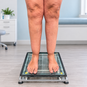
Regardless of its origin, superficial venous insufficiency causes blood to flow backwards, creating an abnormal retrograde circuit and overloading the venous system, which must cope with both normal blood flow and reflux. Over time, this leads to degeneration and dilatation (visible varicose veins) and symptoms such as heaviness, swelling, fatigue and cramps. Here we focus on current treatment options for superficial venous insufficiency.
Conservative treatment
As the name suggests, conservative treatment aims to treat or relieve symptoms of CVI non-invasively. It is indicated for patients who are not suitable for intervention or prefer not to undergo it, those awaiting a procedure and those whose symptoms persist after treatment.
Physical exercise and weight loss are important for reducing symptoms and slowing progression of venous disease.
The use of compression therapy remains the cornerstone of conservative treatment. It has been shown to be effective in controlling symptoms and reducing swelling (oedema). It may also help reduce skin induration in patients with trophic changes (lipodermatosclerosis, atrophie blanche and stasis dermatitis) and is fundamental to healing venous leg ulcers.
Drug treatment has been used for decades, but there is some controversy about its precise role in treating CVI. Venoactive drugs are widely prescribed in certain countries but unavailable in others. Their main mechanisms of action are reducing capillary permeability, reducing the release of inflammatory substances or improving venous tone. They are currently recommended as adjuncts to managing symptoms and oedema in CVI.
Surgical methods
The traditional approach to varicose vein surgery is an established method that has evolved over centuries. Some techniques have remained in use, while others have fallen out of favour. Here I discuss the main techniques that remain useful in selected cases, although we have previously covered this subject in another article.
Saphenous vein stripping
This has historically been the most commonly performed operation for varicose veins. The procedure was described by Mayo and Babcock at the beginning of the twentieth century and has changed relatively little since then.
It involves disconnection of the saphenofemoral junction and complete or partial removal of the great saphenous vein.

Today, with the development of endovenous techniques and minimally invasive surgery, this procedure is increasingly reserved for the uncommon cases in which the great saphenous vein is unsuitable for a less invasive procedure (very dilated, tortuous or containing scar tissue).
Crossectomy or high ligation of the saphenofemoral junction
This consists of disconnecting the saphenofemoral junction without removing the great saphenous vein.
Although this less invasive operation is attractive, recurrence rates have been shown to be higher because the great saphenous vein remains in place. Nevertheless, crossectomy may be useful in selected patients with high surgical and bleeding risk.
Perforator vein ligation
Perforator vein ligation is usually performed alongside other procedures to treat reflux from the deep to the superficial venous system when this occurs in a particular patient. It is particularly useful in advanced venous insufficiency with ulcers or marked trophic changes in the subcutaneous tissues (haemosiderin pigmentation, lipodermatosclerosis or atrophie blanche).
It involves disconnecting one or more incompetent perforator veins in the lower leg or thigh that allow backward blood flow from the deep to the superficial system, increasing venous hypertension in an already overloaded system.

The CHIVA method
CHIVA, from the French Cure Conservatrice et Hémodynamique de l’Insuffisance Veineuse en Ambulatoire (ambulatory conservative haemodynamic treatment of venous insufficiency) , represents a shift in the approach to interventional varicose vein treatment. It is based on preserving the venous network by redirecting abnormal flow without removing the main veins, aiming for a well-drained system without pathological reflux.
To achieve this, CHIVA is based on 4 principles:
- Fragmentation of the hydrostatic pressure column
- Interruption of veno-venous connections
- Preservation of drainage perforator veins
- Removal of inadequately drained varicose tributaries.
The advantages of this technique include minimal invasiveness, less postoperative pain and a faster return to normal activities. Its limitation is that, in a system with multiple connections, it relies heavily on excellent venous mapping by duplex ultrasound.

Endovenous methods
Sclerotherapy
We have previously discussed sclerotherapy for varicose veins in an article explaining the procedure, its main benefits and complications, but will briefly cover it again here.
It involves injecting a sclerosant into varicose veins to damage the vessel wall through an inflammatory response. These agents irritate the venous wall, resulting in its destruction and subsequent obliteration.
Foam or microfoam sclerotherapy and liquid sclerotherapy share the same mechanism of action. The difference is that in foam sclerotherapy, blood within the vein is displaced by the injected foam. This increases the duration of contact between the sclerosant and the inner vessel wall, improving effectiveness.
This technique has important advantages over conventional surgery in selected cases, such as recurrent varicose veins with an irregular distribution, multiple sources of reflux and anatomy that is difficult to treat effectively with surgery. Its minimally invasive nature is also an advantage over other techniques. However, sclerotherapy requires a longer period of follow-up and treatment (in almost all cases, several sessions), often over several months.

Endovenous laser or radiofrequency ablation
Endovenous techniques have undoubtedly transformed phlebology. Thermal ablation of the saphenous vein, whether by endovenous laser ablation or radiofrequency ablation, was developed to offer patients a less painful and less invasive alternative to surgical stripping. We discussed this in a recent article, which you can read here.
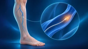
Treatment works by damaging the affected vein (great or small saphenous vein) through direct application of heat to its inner wall, whether from laser energy or radiofrequency energy. Both techniques irreversibly damage the treated vein, resulting in occlusion.
The advantages over stripping are clear in terms of reduced invasiveness, pain and local or systemic complications, and much faster recovery and return to daily activities. Thermal ablation therefore currently has a strong recommendation and high level of evidence for patients with suitable anatomy.

Mechanochemical ablation
Mechanochemical ablation (MOCA) combines sclerotherapy with mechanical injury to the inner wall of the saphenous vein. This relatively new technique further simplifies endovenous treatment compared with laser or radiofrequency ablation, as it does not require tumescent anaesthesia (surrounding the treated vein with anaesthetic solution).
Instead of thermal energy, an intravascular catheter produces mechanical injury either through rotation of its tip or through small elements that scrape the inner vein wall. While the catheter is operated, the surgeon injects a sclerosant, producing additional chemical injury to the vein wall.
Although long-term outcomes for this treatment are not yet available, some studies indicate that results at 3 years are inferior to those of thermal ablation.


Cyanoacrylate adhesive closure
This is a relatively recent method and is not without controversy. Nevertheless, published series report outcomes that are not inferior to thermal ablation. Like MOCA, it is non-thermal and non-tumescent, involving ultrasound-guided delivery of cyanoacrylate into the target vein via an endovenous catheter, using an access technique similar to other endovenous procedures.
Cyanoacrylate rapidly solidifies through polymerisation and induces an inflammatory response in the vein wall. Together with external compression by the surgeon, the adhesive brings the vein walls together and closes the treated vein. At least three types of cyanoacrylate adhesive devices are commercially available for incompetent superficial veins. The main differences concern the formulation and application technique.
As this procedure leaves a foreign material (cyanoacrylate) in the patient's body indefinitely, it remains the subject of substantial debate in medical literature and conferences. In certain cases, significant complications such as local or systemic allergic reactions, including delayed reactions, may limit its use.

So, what is the best treatment for varicose veins?
When faced with a patient who has undergone multiple previous procedures, with difficult anatomy, extensive scarring, recurrent varicose veins and significant venous hypertension, it can be difficult to choose a single optimal technique — because none exists for every case.
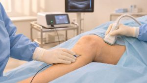
Knowledge and mastery of the available techniques, together with an understanding of what has and has not worked historically, are essential to successful treatment. It is equally important to be skilled in performing and interpreting a duplex ultrasound scan , without which an accurate diagnosis often remains a matter of guesswork.
Every case is therefore different. There is no universally best treatment, but rather the most appropriate treatment and strategy for each individual. Taking all these considerations into account, the key is to find an experienced surgeon, familiar with all the techniques, proficient in duplex ultrasound and able to involve patients actively in their own treatment plan.