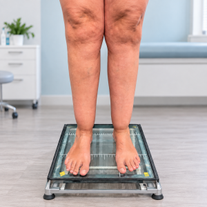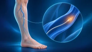
Varicose veins are enlarged veins resulting from structural defects in their walls, which can cause the valves that normally prevent blood from flowing backwards to fail. When discussing varicose veins in the lower limbs, another force must be taken into account: gravity. Blood has to travel upwards against gravity to the heart. When the venous valves fail, blood pools and the veins gradually become enlarged and twisted, forming varicose veins. Understanding this is fundamental to conventional varicose vein surgery.
But how can a closed, enlarged, tortuous vascular system with multiple branches and connections to deeper veins be corrected surgically?
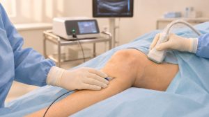
The first step is to understand normal venous anatomy and assess every patient individually. Each person is different and may therefore require a different treatment approach. Physical examination and a duplex ultrasound scan make it possible to map and understand the venous system and plan an intervention if needed (Fig. 1).
Types of conventional varicose vein surgery
Of the various treatment options, conventional surgery is perhaps the one patients fear most. Whether because of concerns about pain, complications or anaesthesia, people tend to delay visiting the angiologist or vascular surgeon until they can no longer tolerate their symptoms. Nowadays, preference is given to minimally invasive procedures such as laser treatment, radiofrequency ablation or sclerotherapy. These are excellent treatment options and may offer better results than surgery, depending on the case. However, they are not suitable in every situation, and traditional surgical removal of varicose veins, incompetent perforators and venous trunks is still necessary in selected cases. Common types of conventional varicose vein surgery include saphenous vein stripping, phlebectomy and the CHIVA method.
Saphenous vein stripping
Saphenous vein stripping is the classic varicose vein operation. It remains a common procedure in some parts of the world and has historically been the most frequently performed operation for varicose veins. It is the benchmark against which the minimally invasive techniques mentioned above have been compared. The procedure was described by Mayo and Babcock (Fig. 2) at the beginning of the twentieth century and has remained relatively unchanged.
It consists of ligation and division of the saphenofemoral junction and its tributaries, together with complete or partial removal of the great or small saphenous vein (Fig. 3). In my current practice, this technique is reserved for cases where minimally invasive endovenous treatment (laser or radiofrequency ablation) is not feasible because the saphenous vein is excessively large or tortuous and/or there are very large perforators. This physically removes the main pathway of venous reflux causing the varicose veins. Blood flow is consequently redirected through normally functioning pathways, chiefly the deep venous system.
CHIVA method
CHIVA, from the French Cure Conservatrice et Hémodynamique de l’Insuffisance Veineuse en Ambulatoire (ambulatory conservative haemodynamic treatment of venous insufficiency), described by Franceschi in 1988, represents a fundamental change in the approach to treating varicose veins. It is based on the venous hypertension caused by pathological reflux in varicose venous networks.
The technique aims to achieve a well-drained venous system without pathological reflux and consequently without venous hypertension, relieving symptoms. This is achieved by selectively interrupting affected saphenous trunks and their drainage tributaries, redirecting flow and dividing the hydrostatic pressure column while preserving the main saphenous veins (Fig. 4).
The advantages of this technique include minimal invasiveness, less postoperative pain and a faster return to normal activities. Its limitation is that, in a system with multiple connections, it relies heavily on excellent venous mapping by duplex ultrasound.
Ambulatory phlebectomy
The term “phlebectomy”, per se, means removal of a vein. In practice, it refers to the technique described by Robert Müller in the 1950s, which involves removing varicose tributaries through small 2–3 mm skin incisions along the enlarged veins. The vein is then hooked with a surgical hook and removed with forceps (Fig. 5).
The incisions are very small and usually require no sutures. Patients recover and are discharged on the same day, as the procedure can be performed under local or regional anaesthesia.
This technique may be used alone when the saphenous trunks and perforator veins are unaffected, or in combination with the other procedures described above, including laser ablation, radiofrequency ablation, foam sclerotherapy and CHIVA.
Conclusion
Surgical treatment of varicose veins remains necessary in many individual cases, and it is the responsibility of the specialist vascular surgeon with expertise in the venous system to devise a surgical strategy tailored to each patient. The choice of technique is not made in the operating theatre, but many days or weeks beforehand, during the consultation and ultrasound assessment. Following diagnostic evaluation, the treatment plan is developed together with the patient once they have been informed about the nature of their condition.
In most cases, the approach is non-surgical, but this depends on each patient's venous system. A combination of techniques is often needed for an individual patient. This greatly increases the vascular surgeon's ability to address the problem and allows a quicker recovery with fewer complications.