The origins of duplex ultrasound
Duplex ultrasound stands out as one of the most important tools in the daily practice of angiology and vascular surgery. It is so integral to diagnosis in our specialty that it is now difficult to imagine a specialist in Angiology and Vascular Surgery who has not mastered its use and particular features.
One notable chapter in the history of duplex ultrasound is the work of the Italian scientist Lazzaro Spallanzani around 1793. Spallanzani studied how various nocturnal animals could fly and find their way in the dark. He established that owls could navigate using their large eyes but could not move around in complete darkness. Bats, by contrast, could fly without difficulty in absolute darkness.
The experiment
In an experiment, he blinded several of these animals and observed that, despite losing their sight, they could still move around and thrive normally. When dissected, their stomachs contained similar quantities of captured insects to those of bats that had retained their sight. When he blocked their ear canals with tiny brass tubes, however, their remarkable ability to navigate at night deteriorated dramatically: they repeatedly collided with any obstacle in their path.
A sixth sense
Spallanzani then suggested that bats might possess a “sixth sense” other than sight, “del quale essendo noi privi, non potremo mai farne idea” (as we lack this sense or organ, I conclude that we can never imagine what it is like), as he wrote in 1794. “Spallanzani's bat problem” would not be fully resolved until much later, in the mid-1930s, when researchers established that bats navigated in flight using high-frequency sounds inaudible to humans, emitted through the mouth and detected by the ears as echoes reflected by obstacles. Through Spallanzani's work, ultrasound first became a subject of human scientific curiosity.

The technique is based on ultrasound. Its introduction as Doppler sonography was made possible by the discovery of the Austrian physicist Christian Doppler (1803-1853), who in 1842 described what we now call the Doppler effect: a change in the frequency of sound waves or electromagnetic radiation when the source is moving relative to an observer. Its medical use is fundamental to the haemodynamic assessment of blood flow.


Over the centuries, as technology advanced, further discoveries led to the development of what we now know as the duplex ultrasound scanner. Its development is undoubtedly one of the most outstanding examples of cooperation across disciplines, sciences, individuals and nationalities in the history of medicine. Vascular duplex ultrasound provides more than an excellent diagnostic tool: it also assists with planning and quality assessment of vascular surgical repairs. It brings a new dimension to the study of vascular physiology and pathology: the ability to assess, compare and quantify. This is a defining feature of science.
Like any diagnostic tool, vascular duplex ultrasound has advantages, disadvantages and limitations. Although I consider the limitations to be few, it is worth knowing what they are.

Benefits of duplex ultrasound
Ultrasound scanners do not require a substantial financial investment, and maintenance is straightforward and inexpensive. Developing the necessary scanning skills is an ongoing process. For a medical specialist, acquiring these skills need not take excessively long. Training can be undertaken safely for patients and is accessible to practitioners with varying levels of technical skill.
Haemodynamic information
Duplex ultrasound makes it possible to describe anatomy in detail, take measurements and even characterise tissue texture and properties. The anatomical information obtained matches or, in some cases, surpasses that of conventional ultrasound.
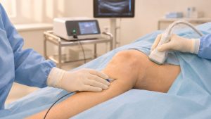
The feature that has brought duplex ultrasound to the forefront of vascular diagnosis is its ability to provide haemodynamic information. Duplex imaging combines Doppler measurements of blood flow with conventional ultrasound images. This allows the effects of a particular abnormality on blood flow to be quantified and helps guide the treatment strategy.


Duplex ultrasound images showing areas of narrowing that restrict blood flow.
No contraindications
Another major advantage of duplex ultrasound is the absence of absolute or relative contraindications to the examination. There is no need to investigate the patient's medical history before deciding to perform the scan, except for information needed to guide the diagnostic examination appropriately. There are no paradoxical or allergic reactions.
Safety of duplex ultrasound
The safety of ultrasound in humans has been widely demonstrated at exposure levels much higher than those used in routine examinations. Ultrasound is in fact used therapeutically for certain conditions, and prenatal ultrasound is widely used with a well-established safety record.
24/7 availability
When integrated into the daily practice of a well-organised vascular surgery department, duplex ultrasound can be available 24 hours a day, every day of the year. We also know that very little equipment is required for these examinations: simply an ultrasound scanner with a couple of probes of different frequencies. An operating theatre or radiology suite is not essential, nor do we necessarily need technical staff to assist us or a fixed timetable.
Versatility
The increasingly compact design of modern ultrasound scanners and the introduction of portable devices have transformed scanning in modern vascular laboratories. Almost any room in a healthcare facility can become a suitable place to work. For example, we can take the ultrasound scanner to the patient's bedside, the operating theatre, intensive care or the emergency department.
Independence
Vascular laboratories that form an integral part of vascular surgery departments allow flexible working without excessive reliance on other departments for diagnosis and treatment planning. This saves time, makes organisation easier and helps optimise resources.
Applications in minimally invasive surgical procedures
As discussed, duplex ultrasound is no longer confined to the vascular examination room. It is also used in the operating theatre to guide surgery and interventions, helping make certain procedures safer, more effective and minimally invasive.
Limitations
Operator dependence
Probably the main limitation of duplex ultrasound is its operator dependence. At least in theory, two clinicians examining the same patient may arrive at different findings.
Various approaches have been developed to minimise this subjective component. Considerable efforts have been made to establish objective criteria for diagnosis, particularly numerical values obtained from Doppler measurements.
Although this is a limitation of the test, it need not be a barrier to its practical use. One of the most important aspects of Doppler examinations is their role in guiding treatment. In practice, developing a reliable diagnostic strategy or treatment plan can be more important than obtaining an exact anatomical diagnosis. Deciding whether a patient is a candidate for surgical or endovascular revascularisation or conservative treatment does not depend solely on a particular numerical reading. Instead, we combine the different parameters we observe (anatomy, greyscale images, colour, velocity waveforms and audible signals) with clinical knowledge and experience. Differences between two duplex ultrasound scans should therefore not automatically be a cause for concern.
Examination time and patient tolerance
Many vascular examinations take considerable time because of the patient's anatomy, difficulties obtaining conclusive images or simply the nature of the examination. In general, Doppler examinations are well tolerated because they are non-invasive and do not cause pain.
Conclusion
Vascular duplex ultrasound and vascular laboratories offer much more than an excellent diagnostic tool. Used effectively and with appropriate follow-up protocols, they are central to evaluating the quality of vascular surgical repairs performed in healthcare centres. They have enabled, and continue to enable, the development of vascular surgery. The vascular surgeon who masters the technique through study and commitment can, like Spallanzani's bat, feel supported in clinical practice by a profound and fascinating sixth sense.